Exploring The Technology Behind Endobronchial Ultrasound
In the realm of modern medicine, technological advancements continually push the boundaries of what is possible in diagnosing and treating various medical conditions. One such innovation making significant strides in the field of respiratory medicine is endobronchial ultrasound (EBUS). This sophisticated technology merges ultrasound imaging with bronchoscopy, offering clinicians a minimally invasive yet highly effective method for visualizing and accessing the structures within the chest, particularly the lungs and surrounding lymph nodes. Let’s delve into the intricacies of this groundbreaking technology and its implications in healthcare.
To Know more about It Please click Here
Understanding Endobronchial Ultrasound (EBUS)
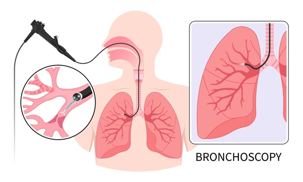
Endobronchial ultrasound (EBUS) is a medical procedure that combines bronchoscopy with ultrasound imaging. It involves the insertion of a bronchoscope equipped with an ultrasound transducer into the patient’s airways, allowing real-time visualization of structures adjacent to the bronchi, such as the lungs and mediastinal lymph nodes. The procedure is commonly used for diagnosing and staging lung cancer, evaluating mediastinal lymphadenopathy, detecting infections, and guiding various therapeutic interventions.
The Technology Behind EBUS
At the heart of EBUS lies ultrasound technology, which utilizes high-frequency sound waves to generate detailed images of internal body structures. In the context of endobronchial ultrasound, a specialized ultrasound transducer is integrated into the bronchoscope. This transducer emits ultrasound waves and receives the echoes bouncing back from surrounding tissues. By analyzing these echoes, sophisticated computer algorithms construct real-time images of the bronchial wall, adjacent lymph nodes, and other structures in the chest.
The EBUS system typically consists of several key components:
Bronchoscope: A flexible, fiber-optic instrument that is inserted into the patient’s airways to provide access to the lungs and surrounding structures.
Ultrasound Transducer: This component is attached to the tip of the bronchoscope. It emits ultrasound waves and captures the returning echoes to generate images.
Ultrasound Processor: A specialized computer system that processes the received ultrasound signals and constructs high-resolution images in real time.
Display Monitor: The generated ultrasound images are displayed in real time on a monitor, allowing the clinician to visualize and interpret the internal structures.
Clinical Applications of EBUS
Endobronchial ultrasound has a wide range of clinical applications, including:
Diagnosis and Staging of Lung Cancer: EBUS allows clinicians to visualize and sample suspicious lesions within the lungs and mediastinum, aiding in the diagnosis and staging of lung cancer.
Evaluation of Mediastinal Lymph Nodes: EBUS enables the assessment of mediastinal lymph nodes for the presence of metastases or other abnormalities, crucial for staging lung cancer and determining treatment strategies.
Detection of Infections: EBUS can help identify infectious processes within the lungs and mediastinum, facilitating targeted diagnostic and therapeutic interventions.
Guidance for Therapeutic Procedures: EBUS guidance is utilized for various therapeutic interventions, such as transbronchial needle aspiration (TBNA), bronchial biopsy, and drainage of mediastinal fluid collections.
Advantages of EBUS
EBUS offers several advantages over traditional diagnostic techniques and surgical procedures:
Minimally Invasive: EBUS is minimally invasive compared to surgical procedures, reducing the risk of complications, postoperative pain, and recovery time for patients.
Real-time Imaging: EBUS provides real-time imaging, allowing clinicians to visualize and target specific structures with precision during procedures.
Accuracy: EBUS offers high diagnostic accuracy in identifying lung lesions, mediastinal lymphadenopathy, and other abnormalities, leading to improved patient outcomes.
Versatility: EBUS is a versatile tool that can be used for both diagnostic and therapeutic purposes, offering clinicians a comprehensive approach to managing respiratory conditions.
Conclusion
Endobronchial ultrasound (EBUS) represents a significant advancement in respiratory medicine, offering clinicians a powerful tool for diagnosing and managing various pulmonary conditions. By integrating ultrasound imaging with bronchoscopy, EBUS provides real-time visualization of internal structures with high diagnostic accuracy while minimizing invasiveness and patient discomfort. In the field of respiratory medicine, EBUS is expected to become more and more important in terms of enhancing patient care and results as technology advances.


