
Exploring the Depths of Diagnosis Understanding Endobronchial Ultrasound
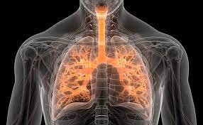
Endobronchial ultrasound (EBUS) is a cutting-edge medical technology that has revolutionized the field of respiratory medicine. This minimally invasive procedure combines bronchoscopy with ultrasound imaging to provide detailed visualization of the airways and adjacent structures within the chest. In this article, we will delve into the intricacies of endobronchial ultrasound, its applications, and the significant impact it has had on the diagnosis and management of respiratory conditions.
The Basics of Endobronchial Ultrasound
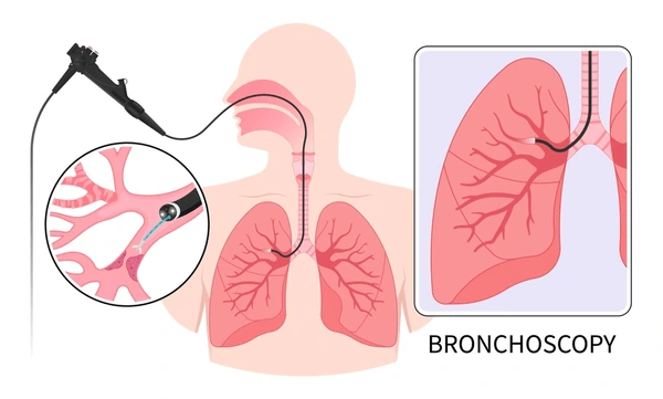
- Procedure Overview: Endobronchial ultrasound involves the insertion of a thin, flexible tube called a bronchoscope through the patient’s mouth or nose and into the airways. The bronchoscope is equipped with an ultrasound transducer at its tip, allowing real-time imaging of the bronchial walls, surrounding lymph nodes, and structures within the chest.
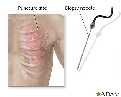
- Real-Time Imaging: One of the key advantages of EBUS is its ability to provide dynamic, real-time ultrasound imaging during the procedure. This allows physicians to visualize abnormalities, take targeted biopsies, and guide various diagnostic and therapeutic interventions.
Applications of Endobronchial Ultrasound
- Lung Cancer Diagnosis: EBUS plays a crucial role in the diagnosis and staging of lung cancer. By allowing physicians to access and sample lymph nodes within the chest, EBUS aids in determining the extent of the disease and guiding appropriate treatment decisions.
- Evaluation of Mediastinal and Hilar Lymph Nodes: EBUS is particularly valuable in assessing lymph nodes in the mediastinum and hilar regions. This is essential for accurate cancer staging, as the presence of cancer cells in these nodes may influence treatment plans.
- Diagnosis of Infections: EBUS is utilized in the diagnosis of infectious diseases affecting the respiratory system, such as tuberculosis or fungal infections. The ability to obtain precise samples enables healthcare professionals to identify the causative agents and tailor treatment accordingly.
- Evaluation of Benign Conditions: Beyond cancer diagnosis, EBUS is employed in the assessment of benign conditions affecting the airways and surrounding structures. This includes evaluating inflammatory conditions, identifying the cause of unexplained coughs, and determining the nature of pulmonary nodules.
Benefits of Endobronchial Ultrasound
- Minimally Invasive: EBUS is a minimally invasive procedure, significantly reducing the risks and discomfort associated with traditional surgical approaches. Patients typically experience shorter recovery times and less postoperative pain.
- Precise Targeting: The real-time imaging capabilities of EBUS allow for precise targeting of lesions or abnormalities, improving the accuracy of biopsies and reducing the need for more invasive procedures.
- Outpatient Setting: Many EBUS procedures can be performed on an outpatient basis, allowing patients to return home on the same day. This contributes to increased convenience and a more streamlined healthcare experience.
Conclusion
Endobronchial ultrasound has emerged as a powerful diagnostic tool in the realm of respiratory medicine, offering healthcare professionals unprecedented insights into the intricacies of the chest and airways. With its ability to facilitate accurate cancer staging, guide therapeutic interventions, and diagnose a range of respiratory conditions, EBUS stands at the forefront of advancing patient care. As technology continues to evolve, the role of endobronchial ultrasound in improving diagnostic precision and treatment outcomes is expected to expand further, shaping the landscape of respiratory medicine for years to come.