Endobronchial Ultrasound (EBUS) in Gurugram for Lung Diagnosis
Endobronchial Ultrasound in Gurugram is an advanced bronchoscopic technique used to evaluate the lungs, airways, and nearby lymph nodes. It combines bronchoscopy with ultrasound imaging and can help doctors obtain tissue samples from areas that may not be accessible through conventional bronchoscopy. EBUS is especially valuable when investigating suspicious lung lesions or enlarged mediastinal and hilar lymph nodes.
What Is Endobronchial Ultrasound?
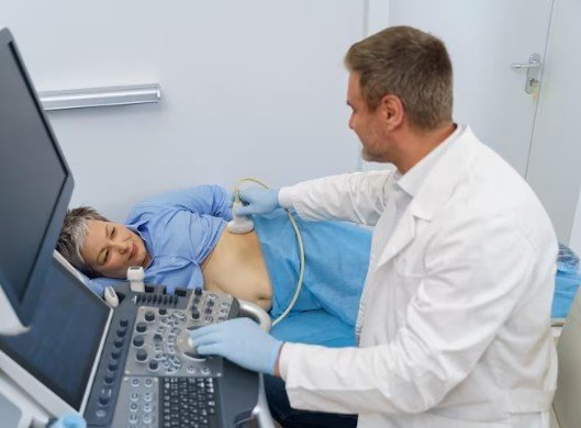
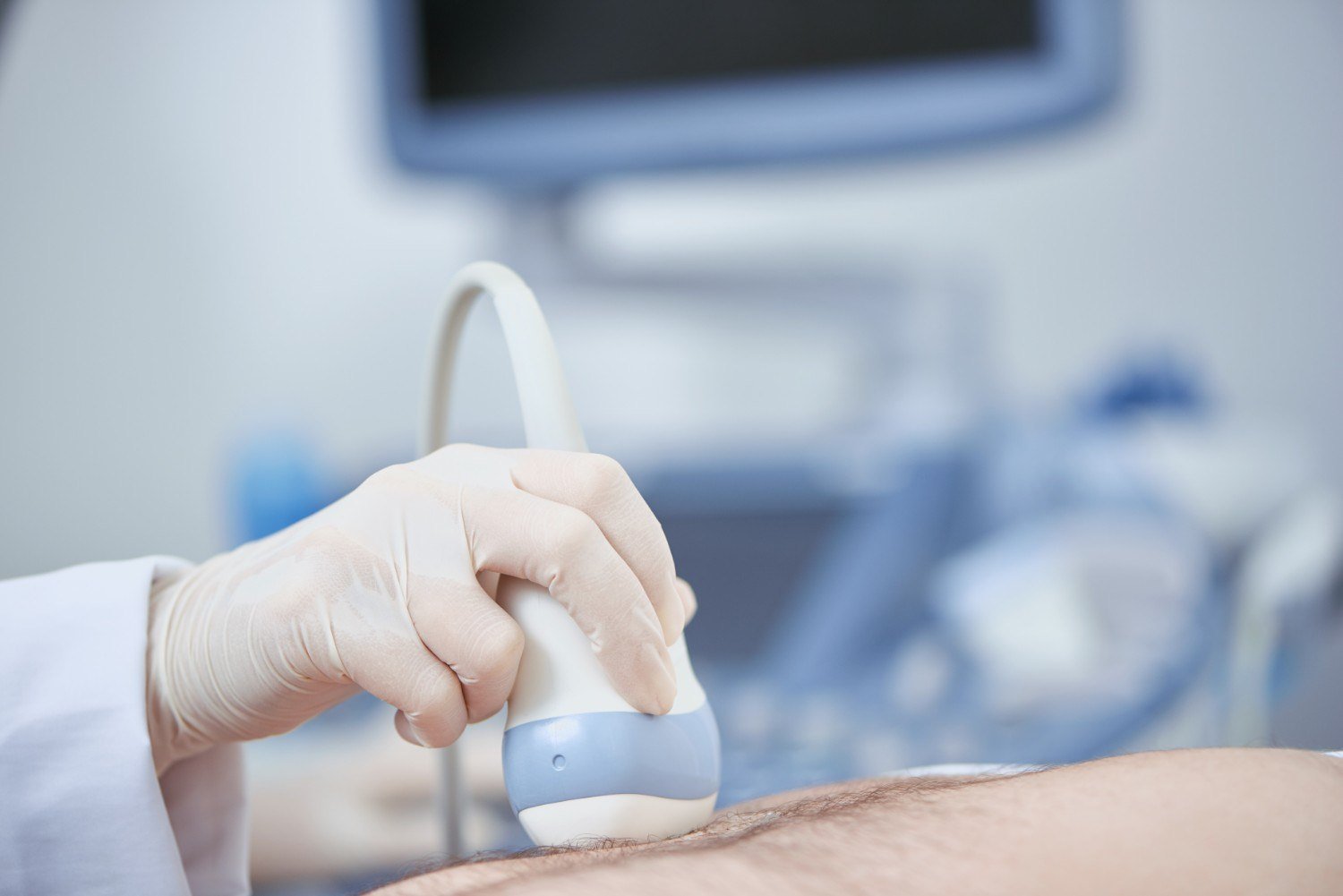
Endobronchial ultrasound, commonly called EBUS, is a minimally invasive diagnostic procedure performed using a specialized bronchoscope with an ultrasound probe. The probe provides real-time images of structures located around the airways.
EBUS can help identify lymph nodes and other abnormalities near the trachea and bronchi. Once the target is located, a fine needle can be guided through the airway wall to collect a tissue or cell sample.
The sample is then examined by a pathologist to help establish the diagnosis.
For fast information, chat directly with us on WhatsApp.
When Is Endobronchial Ultrasound in Gurugram Recommended?
Endobronchial Ultrasound in Gurugram may be recommended when imaging studies such as CT or PET-CT show suspicious lymph nodes or lesions around the airways.
It may be useful for:
- Evaluating enlarged mediastinal lymph nodes
- Investigating suspicious hilar lymph nodes
- Diagnosing selected centrally located lung tumours
- Staging suspected or confirmed lung cancer
- Obtaining tissue for pathological examination
- Assessing certain mediastinal abnormalities
Current European guidelines recommend endosonography, including EBUS-TBNA, as a preferred minimally invasive approach for mediastinal nodal tissue staging in appropriate patients with suspected or confirmed non-small cell lung cancer.
How Does EBUS Help Diagnose Lung Disease?
A conventional bronchoscopy allows a doctor to see the inside of the airways. However, some lymph nodes and abnormalities are located outside the airway wall and cannot be directly visualised.
This is where Endobronchial Ultrasound in Gurugram provides an important advantage. Ultrasound imaging helps locate structures beyond the airway wall. A fine needle can then be guided to the selected lymph node or lesion.
The collected material can be used for cytological or histopathological assessment. In patients with suspected lung cancer, EBUS-TBNA can also provide tissue that may assist with treatment planning and biomarker assessment.
EBUS for Lung Cancer Diagnosis and Staging
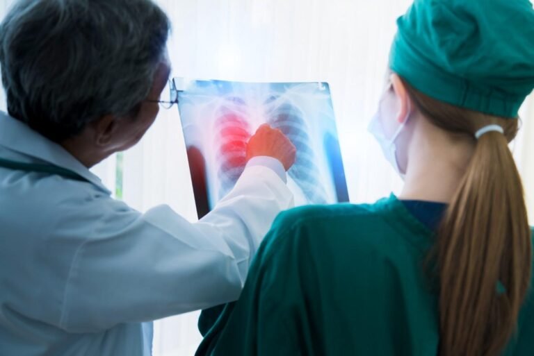
Accurate staging is essential when lung cancer is suspected or confirmed. It helps determine how far the disease has spread and supports treatment planning.
EBUS-TBNA is particularly useful for sampling mediastinal and hilar lymph nodes. Current guidance considers systematic EBUS-TBNA an important standard for mediastinal nodal staging, while combining EBUS with oesophageal ultrasound-based sampling may improve access to additional lymph node stations in selected cases.
Therefore, Endobronchial Ultrasound in Gurugram can play an important role in obtaining diagnostic information without immediately requiring a surgical staging procedure.
What Happens During an EBUS Procedure?
Before the procedure, the doctor reviews the patient’s symptoms, imaging scans, medical history, and overall health.
During EBUS, a flexible bronchoscope is introduced through the mouth or nose and guided into the airways. The ultrasound probe is then used to identify the target lymph node or lesion.
If tissue sampling is required, a fine needle is passed through the airway wall under real-time ultrasound guidance. Multiple samples may be collected for laboratory analysis.
The procedure is generally performed with appropriate sedation or anaesthesia. Afterward, the patient is monitored until the effects of sedation have reduced.
Benefits of Endobronchial Ultrasound in Gurugram
There are several reasons why Endobronchial Ultrasound in Gurugram is increasingly used for selected pulmonary and thoracic conditions.
Key benefits include:
- Minimally invasive tissue sampling
- Real-time ultrasound guidance
- Access to lymph nodes around the airways
- Useful for lung cancer staging
- May reduce the need for more invasive diagnostic procedures
- Can provide samples for pathological evaluation
- Helps support treatment planning
The latest ERS/ESGE/ESTS guidance recommends endosonography over mediastinoscopy for appropriate mediastinal nodal staging, reflecting its role as a less invasive diagnostic and staging approach.
Risks and Possible Complications
Although EBUS is minimally invasive, it can still have risks. Possible complications include temporary throat discomfort, cough, minor bleeding, infection, oxygen desaturation, and, less commonly, more significant bleeding or other complications.
The individual risk depends on the patient’s medical condition, the location being sampled, and the complexity of the procedure. Careful assessment and appropriate procedural expertise are important for maintaining safety.
Best EBUS Doctor in Gurugram – Dr. Bharat Gopal
Choosing the Best EBUS Doctor in Gurugram is important when advanced bronchoscopic evaluation or lymph node sampling is required. Dr. Bharat Gopal evaluates each patient based on their symptoms, chest imaging, medical history, and suspected lung condition before recommending EBUS. The procedure may be considered for evaluating enlarged mediastinal or hilar lymph nodes, obtaining tissue samples, and supporting the diagnosis and staging of selected lung diseases. A careful pre-procedure assessment also helps determine the appropriate sampling approach and ensures that the potential benefits and risks are discussed with the patient.
Who May Need an EBUS Evaluation?
Not every patient with a lung nodule, cough, or enlarged lymph node requires EBUS. The procedure is considered based on imaging findings, suspected diagnosis, symptoms, and whether obtaining tissue would change management.
A pulmonologist or interventional pulmonology specialist may recommend Endobronchial Ultrasound in Gurugram when tissue sampling or lymph node staging is clinically important.
Conclusion
Endobronchial Ultrasound in Gurugram is an advanced minimally invasive technique that can assist in the diagnosis and staging of selected lung conditions. By combining bronchoscopy with real-time ultrasound and needle sampling, EBUS allows doctors to evaluate lymph nodes and lesions located around the airways.
For patients with suspicious lung findings or enlarged mediastinal lymph nodes, appropriate specialist evaluation can determine whether EBUS is suitable and whether the information obtained can help guide further treatment.
Frequently Asked Questions
1. What is EBUS used for?
EBUS is primarily used to evaluate structures around the airways, obtain tissue samples from selected lymph nodes or lesions, and assist with lung cancer diagnosis and staging.
2. Is EBUS a surgical procedure?
No. EBUS is performed through a flexible bronchoscope and is considered a minimally invasive bronchoscopic procedure.
3. Can EBUS diagnose lung cancer?
EBUS can help obtain tissue or cell samples from appropriate lung lesions and lymph nodes. The final diagnosis is made after laboratory and pathological evaluation.
4. Is EBUS useful for lymph node staging?
Yes. EBUS-TBNA is widely used for sampling mediastinal and hilar lymph nodes and plays an important role in staging selected lung cancer patients.
5. Is Endobronchial Ultrasound suitable for everyone?
No. The need for EBUS depends on the patient’s imaging findings, suspected condition, medical history, and whether tissue sampling is clinically necessary.
For more details or to book an appointment, visit their official website: https://delhichestspecialist.com/.